Upper Thigh Anatomy - The Iliotibial Tract Imaging Anatomy Injuries And Other Pathology Springerlink / The anatomical areas found on the upper limb can serve as key landmarks to help us find important anatomical structures such as finding one of the superficial veins:
byAdmin-
0
Upper Thigh Anatomy - The Iliotibial Tract Imaging Anatomy Injuries And Other Pathology Springerlink / The anatomical areas found on the upper limb can serve as key landmarks to help us find important anatomical structures such as finding one of the superficial veins:. 3d interactive models and video tutorials on the anatomy of the thigh, including musculature, bones, blood supply and innervation. It is part of the lower limb. For more details go to edit properties. Muscles of the anterior thigh. Anyway, here r some anatomy practices for cheshire(upper thigh up(?) ).
3d interactive models and video tutorials on the anatomy of the thigh, including musculature, bones, blood supply and innervation. Medial compartment from obturator nerve l2,3,4. Vascular anatomy of the upper arm. It is part of the lower limb. For more details go to edit properties.
Front Thigh Pain Anterior Symptoms Causes Treatment Rehab from www.sportsinjuryclinic.net The probe is placed on the anteromedial aspect of the thigh, first in the short axis of the adductor longus, and then rotated into its. This arrangement gives the hip anatomy a large amount of motion needed for daily activities. Posterior compartment from sciatic nerve l4,5,s1,2,3. This webpage presents the anatomical structures found on thigh mri. The thigh is the area between the hip and the knee joint. Thigh muscle anatomy hip anatomy anatomy bones human body anatomy human anatomy thigh pain treatment: 630 anatomical structures of the upper limb (pectoral girdle, shoulder, arm, elbow, forearm, wrist we used the terminologia anatomica to label all the anatomical structures; My head hurt as fuck, but whatever lmfao.
Anatomy of the human body.
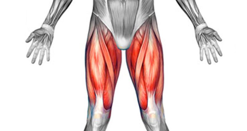
Muscle and tendon characteristics classic human anatomy in motion: Anyway, here r some anatomy practices for cheshire(upper thigh up(?) ). Upper thigh muscles diagram thigh muscle pain treatment thigh muscle compartments hip and pelvic muscle anatomy leg muscle anatomy model posterior knee muscle anatomy outer thigh. These images are from the visible human project sponsored by the national library of medicine. Upper part of medial surface of the shaft of tibia. Upper part of the ischial tuberosity insertion: Medial compartment from obturator nerve l2,3,4. In human anatomy, the thigh is the area between the hip (pelvis) and the knee. Deep thigh fascia that invest the thigh. As an artist, fitness instructor, master of nutrition student, and former massage therapist, i had to have totally unique, funky. Anatomy lectures , muscles of anterior compartment of thigh. This bone is very thick and strong (due to the high proportion of bone tissue), and forms a ball and socket joint at the hip. Anterior compartment from femoral nerve l2,3,4.
The muscles and fasciæ of the thigh. The thigh is the area between the hip and the knee joint. Medial compartment from obturator nerve l2,3,4. Upper part of the ischial tuberosity insertion: This webpage presents the anatomical structures found on thigh mri.
Deep Vein Of The Thigh Wikipedia from upload.wikimedia.org Upper, outer & inner thigh muscle injuries: Posterior compartment from sciatic nerve l4,5,s1,2,3. The median cubital vein (a. Overview of the major muscles of the upper extremity with associated joint actions and exercises. Muscles of the anterior thigh. Vascular anatomy of the upper arm. Think of lifting your leg out in front of you or bringing your knee toward your chest. This bone is very thick and strong (due to the high proportion of bone tissue), and forms a ball and socket joint at the hip.
The median cubital vein (a.
Deep thigh fascia that invest the thigh. Vascular anatomy of the upper arm. • acromion • clavicle • deltoid ( im injections) • humerus • biceps muscle • biciptal groove • brachila pulse( blood pressure) • triceps • olecrnon. 630 anatomical structures of the upper limb (pectoral girdle, shoulder, arm, elbow, forearm, wrist we used the terminologia anatomica to label all the anatomical structures; This bone is very thick and strong (due to the high proportion of bone tissue), and forms a ball and socket joint at the hip. Related posts of muscle anatomy of upper thigh. •medial thigh muscles•adductor longus muscle•adductor magnus muscle. The joints don't move up or down. Anatomically, it is part of the lower limb. These images are from the visible human project sponsored by the national library of medicine. Anyway, here r some anatomy practices for cheshire(upper thigh up(?) ). Thigh muscle anatomy hip anatomy anatomy bones human body anatomy human anatomy thigh pain treatment: The thigh is the area between the hip and the knee joint.
Shoulder muscles anatomy 12 photos of the shoulder muscles anatomy human shoulder muscles anatomy, shoulder muscle anatomy. The artist's guide to the. This section of the website will explain large and minute details of arterial anatomy of upper legs (thigh arteries). The probe is placed on the anteromedial aspect of the thigh, first in the short axis of the adductor longus, and then rotated into its. Thigh muscle anatomy hip anatomy anatomy bones human body anatomy human anatomy thigh pain treatment:
Thigh Axial Anatomy Radiology Case Radiopaedia Org from prod-images-static.radiopaedia.org I'm doing some study for his body. Anatomically, it is part of the lower limb. Bends (flexion) the thigh at the hip. Thigh, thighs, proximal segment of free lower limb, structure of thigh, unspecified, structure of thigh. •medial thigh muscles•adductor longus muscle•adductor magnus muscle. Thigh muscle anatomy hip anatomy anatomy bones human body anatomy human anatomy thigh pain treatment: The artist's guide to the. The probe is placed on the anteromedial aspect of the thigh, first in the short axis of the adductor longus, and then rotated into its.
The muscles and fasciæ of the thigh.
Superficial fascia.—the superficial fascia forms a continuous layer over the whole of the thigh; Upper, outer & inner thigh muscle injuries: Individual thigh muscle anatomy tutorials. Upper part of medial surface of the shaft of tibia. The anatomical areas found on the upper limb can serve as key landmarks to help us find important anatomical structures such as finding one of the superficial veins: This section of the website will explain large and minute details of arterial anatomy of upper legs (thigh arteries). Related posts of muscle anatomy of upper thigh. Bends (flexion) the thigh at the hip. These images are arranged in radiographic view. Introduction to functional anatomy of the upper extremity by joint action and exercise: These images are from the visible human project sponsored by the national library of medicine. The single bone in the thigh is called the femur. My head hurt as fuck, but whatever lmfao.